
Step 1: The roots of a skin cancer may extend beyond what is visible on the surface. Mohs surgery is designed to identify and remove these extensions. | 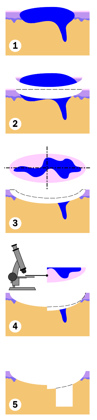 |
Step 2: A layer of skin is removed surgically with a 1-2 mm margin. | |
Step 3: The physician divides the tissue into sections and then color codes the sections with dye. Reference marks are made on the skin to orient the sections. A map of the surgical site is then made. | |
Step 4: The entire base of the specimen is examined microscopically for evidence of remaining cancer. If remaining cancer cells are found, another layer of skin will be removed precisely where cells were seen according to the map. | |
Step 5: Once there is no longer evidence of cancer cells, a surgical reconstruction of the site is completed. |
Mohs micrographic surgery is a specialized, staged technique used to treat selected skin cancers. The surgeon removes a thin layer of tissue, maps and examines the complete surgical margin under a microscope, and removes additional tissue only where cancer remains. This approach provides a high cure rate while preserving as much healthy tissue as possible.
During each stage, your surgeon removes a narrow layer of tissue around and beneath the visible tumor or biopsy site. The tissue is carefully mapped, color-coded, frozen, cut into thin sections, and prepared on microscope slides in our laboratory. Your surgeon examines the complete margin. If cancer cells remain, the map identifies their location so another thin layer can be removed precisely from that area. These steps are repeated until the examined margins are clear. Cure rates vary with the cancer type, location, size, and whether it has been treated previously; your surgeon will discuss what is expected in your individual case.
After the cancer is removed, your surgeon will recommend the repair that best fits the size, depth, and location of the wound. Options may include a side-to-side closure with stitches, a skin flap, a skin graft, or allowing the area to heal naturally. Occasionally, a repair is completed in stages or coordinated with another specialist. Our team will provide written wound-care instructions and arrange follow-up based on your procedure.
Infrequently, a tumor may turn out to be much larger than anticipated. Under these circumstances, reconstruction may require another specialist. This surgery may take place on the same day or on a subsequent day. It is very rare that reconstruction may require hospitalization.
Basal cell carcinoma and squamous cell carcinoma are the skin cancers most often treated with Mohs surgery. Their microscopic extensions may reach beyond what can be seen on the skin. Mohs surgery is especially useful when a tumor is located in a tissue-sensitive area, has aggressive features, has returned after previous treatment, or has poorly defined borders. Your referring clinician and Mohs surgeon determine whether this technique is appropriate for your diagnosis.
Melanoma and other less common tumors may require a different surgical approach, specialized margin testing, delayed repair, or coordination with other specialists. Treatment is individualized according to the pathology diagnosis and current clinical guidance. Your surgeon will explain the recommended plan before your procedure.